
Hello!
I am Dr. Hyuk-chan Cho, Director of Cleor Clinic Uijeongbu Branch.
After receiving laser treatments,
some people say things like this:
"I definitely received a pigment laser treatment,
but my melasma is still the same."
"I've had several pore treatments,
why am I the only one not seeing results?"
When results fall short of expectations, people often blame the equipment or the clinic first,
but in many cases, the cause lies elsewhere.
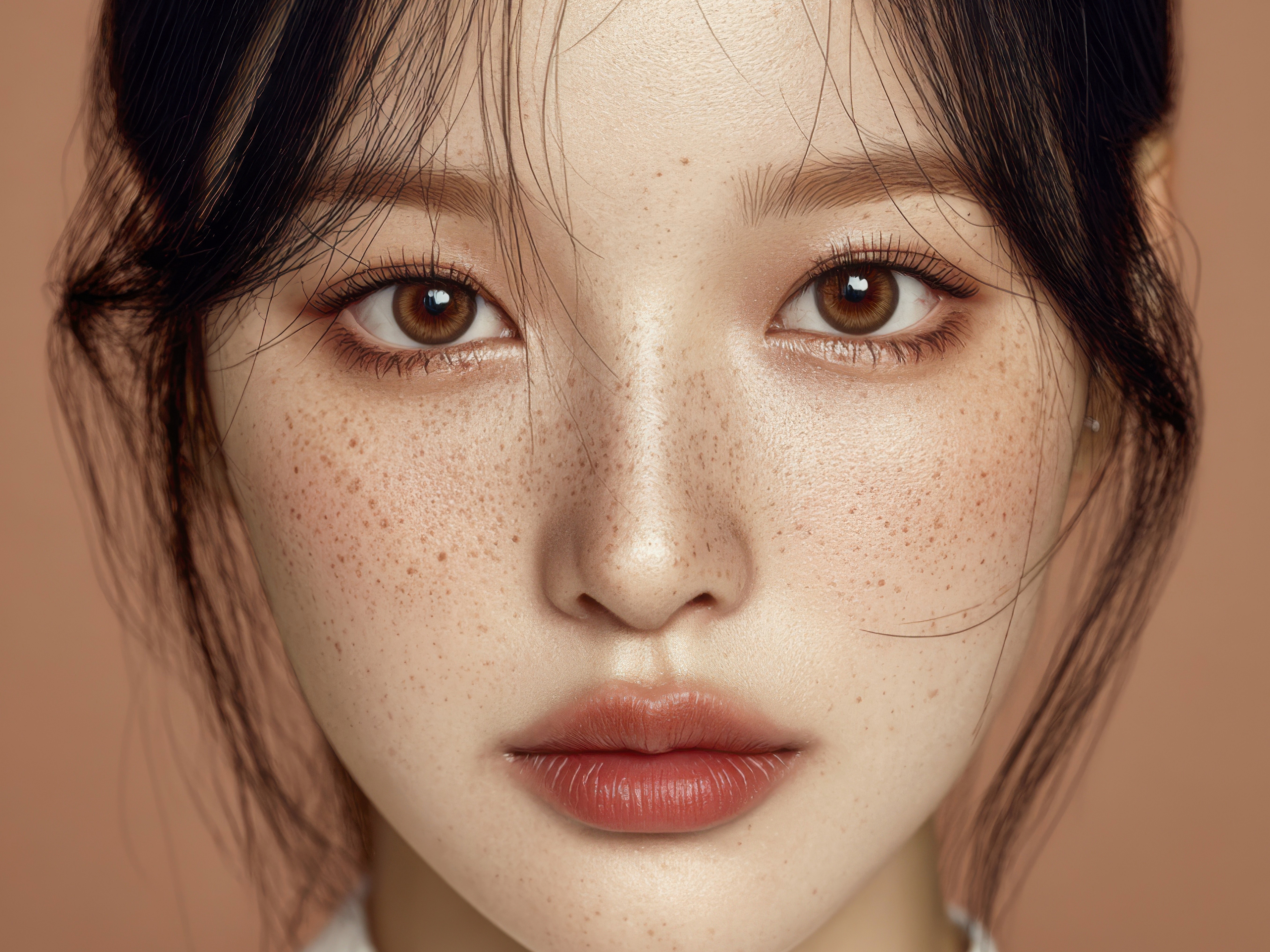
It is because your skin condition
was not accurately identifiedbefore the procedure.
You might have received melasma treatment because it looked like melasma,
but it was actually blemishes, or you thought it was epidermal pigment
when it was actually pigment settled deep within the dermis.
If the starting point is wrong, even the best procedure
will struggle to deliver the expected results.
That is why at Cleor Clinic Uijeongbu Branch,
we first conduct a Mark-Vu skin analysis before any procedure.
Today,I will explain exactly what the Mark-Vu device is,
which procedures it is especially necessaryfor,
and even how it can be utilized after the procedure,
just like a real consultation.
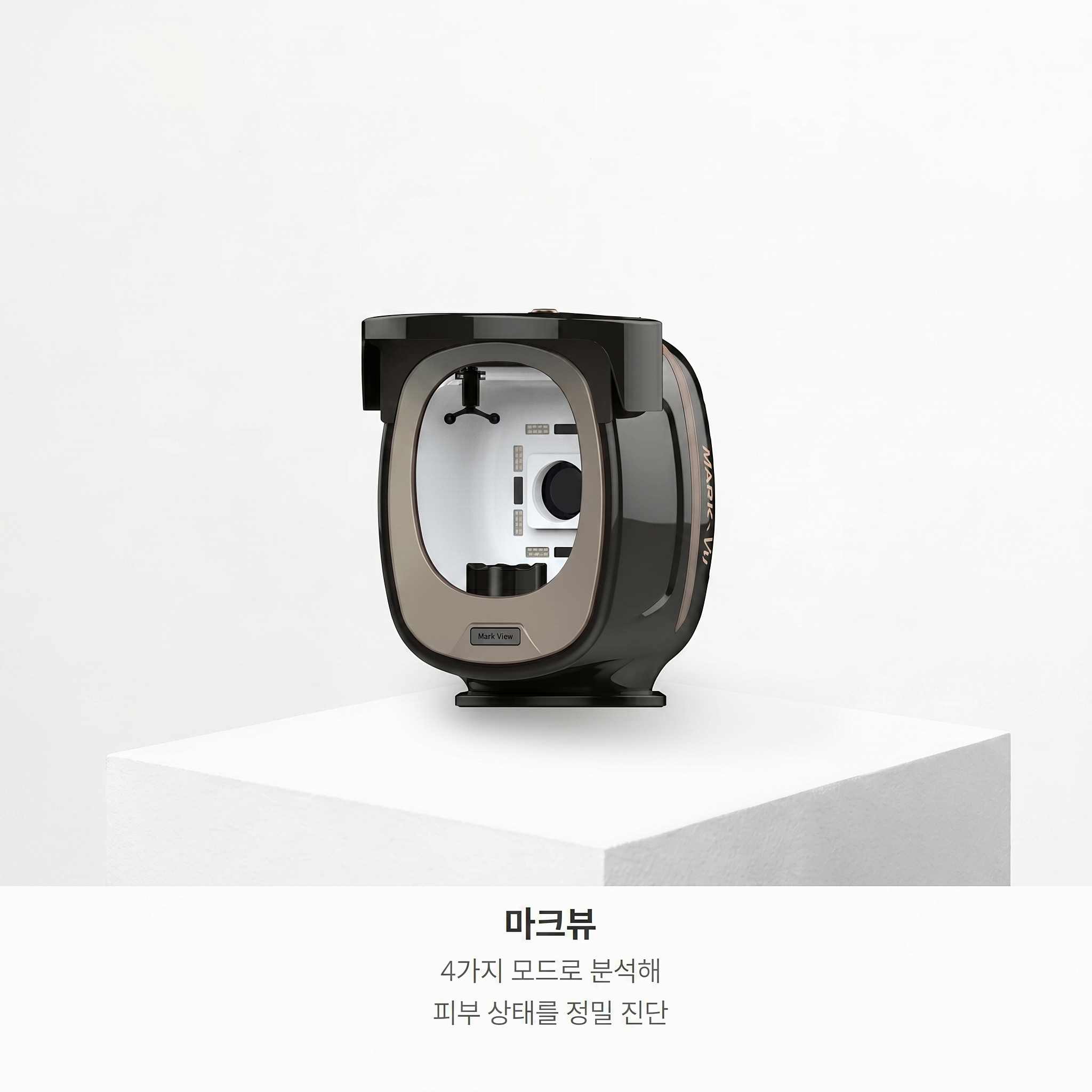
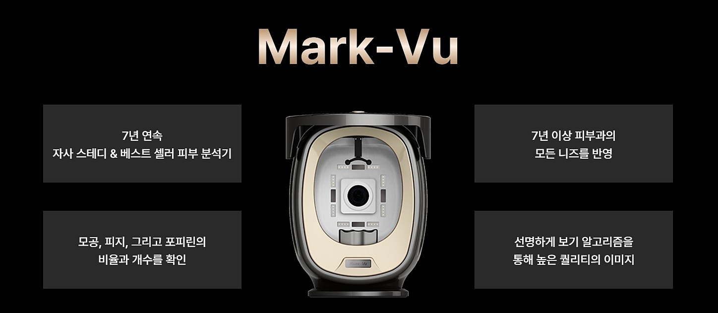
What kind of device is the Mark-Vu?
Mark-Vuis
a skin diagnosis system that uses an 18-megapixel ultra-high-definition camera
combined with four types of LED light sources
to precisely capture and analyze skin conditions.
The biggest difference from standard skin diagnosis equipment
lies in the light source method.
Most skin diagnostic devices use a flash method
that emits a momentary strong light.
Using a flash causes light reflection,
which can make the skin look better than it actually is
or, conversely, create distortions that make issues look exaggerated.
Mark-Vu uses a continuous LED light source.
In simple terms,it captures the skin as it is,
under constant and uniformly distributed light,
much like a natural photo taken under a fluorescent lamp.
In addition, 👉a polarizing filter is added to capture fine details invisible to the naked eye,
such as fine lines, deep-seated pigments,
and the extent of facial flushing.
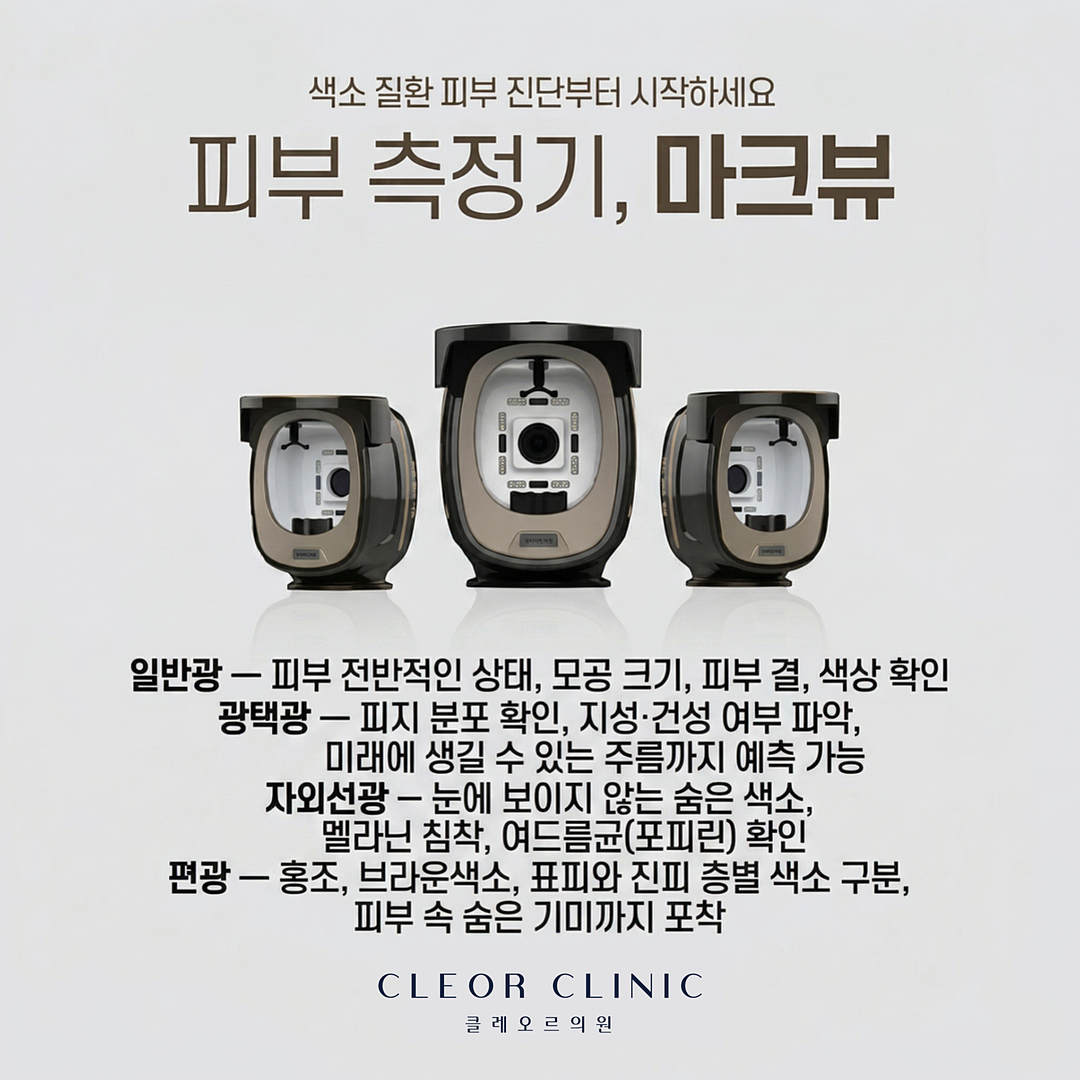
What does each of the four light sources show?
The core of Mark-Vu
is that each light source reveals different skin information.
Normal Light
This is the most basic imaging mode.
It captures the skin in a state closest to what is seen with the naked eye,
checking overall skin condition, pore size,
skin texture, and color.
It is used to record the baseline skin condition
before the procedure.
Specular Light
By maximizing surface reflection,
it highlights sebum distribution and skin irregularities.
It is characterized by its ability to not only identify current skin types
but also predict future wrinkles
that have not yet formed.
UV Light
This mode brings hidden problems that are invisible to the eye
to the surface.
It can identify latent melanin pigment within the skin
and even traces left by acne bacteria (porphyrins).
Polarized Light
This method suppresses surface reflection
to clearly highlight only the pigmentation.
It can distinguish between epidermal and dermal pigments by layer,
and identify the extent of flushing, brown spots, deep dermal melasma,
and dark circles.
By synthesizing these four light sources,
it analyzes more than 12 skin issues at once, including pores, wrinkles, future wrinkles, pigmentation,
melanin, skin tone, dark circles, flushing,
brown spots, sebum, porphyrins, and radiance.
Which procedures is it especially necessary for?
Those considering pigment laser treatments
Even if melasma and blemishes look similar on the surface,
their skin layers and causes are completely different.
Through UV and polarized light analysis,
we must first check whether the pigment is in the epidermis or dermis
and how deep it is located
to select the appropriate laser.
Starting without this process
can lead to being treated with a laser of the wrong wavelength.
Those preparing for lifting procedures
For procedures that address elasticity and sagging, such as Ulthera and Thermage,
identifying the current skin thickness and elasticity
has a significant impact on the treatment design.
The pre-treatment data recorded with Mark-Vu
also serves as a baseline for comparing subsequent changes.
Those considering pore or sebum treatments
The direction of treatment can be decided after objectively confirming
sebum distribution and pore size through specular light analysis.
It is especially useful for those who say, "I think my pores are large,
but I don't know exactly how large they are."
Those with sensitive skin who are unsure which procedures are possible
By first identifying the flushing index and skin sensitivity,
we can more safely determine which procedures
and at what intensity can be performed
based on the current skin condition.
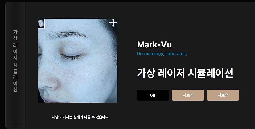
It can also be utilized after the procedure
Another role of Mark-Vu
is the before-and-after comparison.
By saving images taken under identical conditions
by date
and comparing them side-by-side,
it provides more than just a vague feeling of,
"I think something improved, but I'm not sure how much it changed."
You can objectively confirm how pigment density, pore size, and flushing extent
have changed through data and images.
Especially for treatments where changes appear gradually, such as pigment lasers or pore treatments,
the Mark-Vu before-and-after comparison is even more meaningful.
How is the Mark-Vu analysis conducted?
First, after cleansing the skin,
you sit in front of the Mark-Vu device
and images are taken sequentially with the four light sources.
The imaging time is short,
and since there is no direct contact or irritation to the skin,
you can receive it without any burden.
Once imaging is complete, you will review the analysis images and data with the medical staff
to identify which skin concerns should be addressed first
and which procedures are suitable for your current skin condition.
⚠️
Precautions For the most accurate results, Mark-Vu imaging should be done on a cleansed face
without moisturizer, sunscreen, or makeup.
If products remain on the skin, they can affect the light source reaction,
leading to an analysis that differs from the actual skin condition.
Additionally, Mark-Vu is an analytical tool used to understand skin condition
and set the direction for treatment.
The final decision on the procedure based on the analysis results must
be made through a direct consultation with the medical staff.
❓ Frequently Asked Questions
Q. Can anyone receive a Mark-Vu analysis?
A. Yes, everyone from those with skin troubles
to those who are simply curious about their skin condition
can receive it.
It is often conducted for the purpose of understanding the current skin
before deciding on a procedure.
Q. Does it help distinguish between melasma and blemishes?
A. Through UV and polarized light analysis, epidermal and dermal pigments can be identified by layer,
which serves as a reference for identifying pigment types
that are difficult to distinguish with the naked eye.
An accurate diagnosis is made by the medical staff
based on the analysis results.
Q. What are the benefits of receiving Mark-Vu after a procedure?
A. By imaging under the same conditions as before the procedure,
images and data can be compared side-by-side.
It is especially useful for those who want to verify changes
through data rather than just a feeling.
Q. Can I also find out my skin age?
A. You can check reference values for your skin condition
compared to the average for the same age group.
However, this is for reference only,
and a comprehensive judgment is made along with the actual skin condition.
Regardless of the procedure you receive at a dermatology clinic, the first thing needed
is to accurately understand your current skin condition.
Identifying what is needed and where based on data,
rather than just a feeling,
is the first step
toward achieving satisfying results.
At Cleor Clinic Uijeongbu Branch, we will help you identify
what your skin actually needs right now
through Mark-Vu analysis.
Please feel free to contact us for a consultation if you have any questions.
함께 확인하고 안내해드리겠습니다.
궁금한 점은 편하게 상담 문의 주세요.